
num. 7-12 dicembre 2007

back to index
 |
page 002 |
page 004 |
 |
Conchiglie ai raggi X
ClaudioFanelli
ClaudioFanelli
|
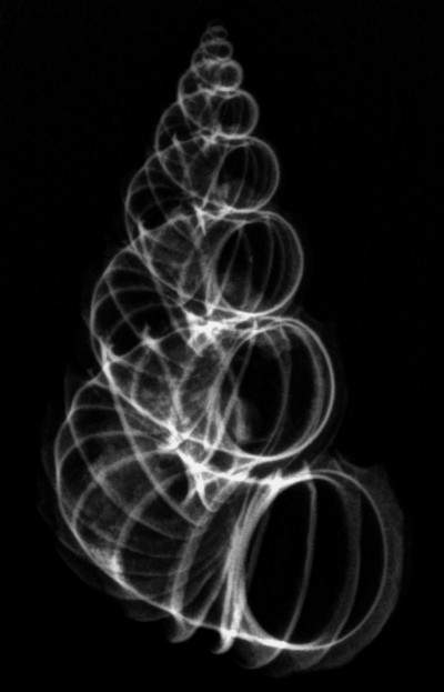
Vi mostro con piacere i risultati raggiunti nel campo della radiografia di conchiglie con mezzi tradizionali, ovvero fotografia su lastre, o pi˘ moderni, usando sensori connessi direttamente ad un computer ed ottenendo quindi direttamente immagini digitali in formato .TIFF. Stiamo parlando ovviamenti di strumenti non disponibili per questo scopo specifico, dato il loro alto costo, ma di strumenti professionali usati nel campo medico. Per chi volesse tentare diciamo subito che l'amicizia con un radiologo, per fare foto su lastre a raggi x, o con un dentista, per l'uso di sensori uniti al computer, Ë fondamentale e comunque non impossibile. Ringrazio a tal proposito due professionisti nel campo che mi hanno permesso di realizzarele immagini di questo articolo: Il prof. Giovacchino Pedicelli, radiologo, e il dott. Fernando Ricci, dentista. Le immagini su lastre sono state ottenute fissando le conchiglie con colla su un foglio di cartone che Ë poi stato radiografato regolando l'intensit‡ del proiettore a raggi x su emissioni deboli, quindi con una potenza molto bassa. Una singola lastra di grande formato ha permesso di fotografare tutte le conchiglie con una sola esposizione. La lastra ottenute Ë poi stata rifotografata con macchina digitale da 6 Mpixel, sia per intero che nei dettagli. Questo passaggio ha degradato, anche se marginalmente, la qualit‡ delle immagini stesse. Queste lastre risalgono a circa 20 anni fa e io non sono quindi in grado di documentare se e quanto la qualit‡ possa essere migliorata in questo lasso di tempo. Di questo fattore si dovr‡ tenere conto nel raffronto che appare in fondo a questa pagina Le immagini ottenute con un sensore collegato direttamente ad un PC sono invece recenti. Le conchiglie sono state di volta in volta posizionate sul sensore che misura circa cm 3x4. Solo in alcuni casi abbiamo potuto fotografare pi˘ di una conchiglia per volta Eccovi quindi un raffronto diretto tra due foto della stessa conchiglia. L'immagine a sinistra Ë stata fatta su lastra mentre quella a destra su sensore. Appare evidente la migliore qualit‡ offerta dal sensore. |
It is my pleasure to show you the results achieved in X-raying shells when using both the traditional methods i.e. X-ray photographs onto negative plates, and the more modern sensors having a direct computer connection and thereby obtaining digital images directly in a TIFF format. Obviously we are talking about instruments not usually utilized for this specific aim, given their prohibitive cost, but about instruments used professionally in the field of medicine. To anyone who would attempt what we have done, we would hastely add that friendship with a radiologist, (to take X-ray photos on plates), or even with a dentist, (to use the sensors linked to a computer), is something fundamental but not impossible. I therefore heartily thank the two professionals who permitted me to use their equipment, thus enabling me to prepare the images for this article: viz Prof. Giovanacchino Pedicelli ñ radiologoist and Dr. Fernando Ricci ñ dentist. The images on X-ray plate were obtained by glueing the shells onto a piece of cardboard which was then X-rayed in the normal way. The intensity of the X-ray projection was regulated to a weak emission and therefore of low intensity. By using only one large photographic plate, we were able to X-ray all the shells in one go. The plate was then re-photographed using a digital machine ñ 6 Mpixel, both for the shell-interiors and for details. However in the act of image transference the quality of the images did degrade somewhat - even if only slightly so. The plates used were those of about 20 years ago and so I cannot truly state whether the quality of plate used today would have been better or not. One will have to judge this by comparison of the images that appear at the bottom of this page. The pictures obtained through the use of a sensor with a direct connection to a computer, are however of recent production. The shells were positioned one by one onto the sensor which measured about cm 3x4. Only in some cases were we able to photograph more than one shell at a time. Here therefore is the direct comparison of two photographs taken of the self-same shell. The one on the left was taken by X-ray onto a plate while that on the right was obtained by using a sensor. The superior quality of the latter (sensor photo), is blatantly apparent. |

|
La mia conclusione Ë che i moderni sensori offrono immagini pi˘ nitide e dettagliate ma non Ë da negare alla foto su lastra il vantaggio di poter fotografare molti esemplari tutti insieme e di poter radiografare anche conchiglie di grandi dimensioni. Nelle foto su sensore noterete una fastidiosa retinatura di fondo che Ë dovuta al sensore che non Ë costituito da una singola cella sensibile ma da pi˘ celle affiancate i cui margini appaiono nell'immagine. Vi prego infine di notare che nelle pagine che seguono, dove di ciascun esemplare viene mostrata l'immagine della conchiglia affiancata a quella a raggi X, non sempre sono riuscito a riprendere il soggetto nella stessa identica posizione. Infine vi lascio una immagine originale, in formato .TIFF per chi voglia esaminarne i dettagli pi˘ piccoli.( 1.879 kb)  La potete vedere da qui o aprirla con il vostro programma preferito cercandola sul Cd con il nome celesti.tif.
|
I have arrived at the conclusion that the modern sensors offer a sharper and more detailed image but at the same time, it cannot also be denied that the X-ray photos have the added advantage of being able to photograph shells of large dimension.
In the photos taken by sensor, you will notice a bothersome network of lines in the background. This is due to the fact that the sensor is composed of not only one sensitive cell, but of several cells placed side-by-side. Hence the margins of these are visible in the resulting image.
Finally please note that where some specimens are shown in the following pages - the images of the shells placed next to those taken by X-ray - I have not always managed to photograph the same specimens in identical positions.
In closing, I leave an original image in TIFF format for all those who wish to examine the specimens in finer detail.( 1.879 kb)
You can see here her from or to open with your preferred program looking for on the Cd with the name celesti.tif.
|
|
Qui a fianco potete vedere l'attrezzatura usata per queste fotografie. Il proiettore di raggi X sul lato destro Ë quello in normale dotazione per le radiografie, mentre nella zona cerchiata in rosso si puÚ vedere il piccolo, ma efficace, sensore della Kodak. Presiede alle operazioni l'assistente Sabrina che, con grande esperienza e capacit‡, ha saputo fornirci gli ottimi risultati mostrati in questo articolo. Occorre tener presente che, mentre le apparecchiature per la radiografia odontotecnica sono gi‡ predisposte per segnali idonei allo spessore e alla consistenza di una conchiglia, quelli per la radiografia corporea debbono essere regolati per bassissime emissioni come se si dovesse, ad esempio, radiografare le dita di una mano.
|
Here on the side you will see the equipment used to obtain the photographs. The X-ray machine on the right is a normal standard machine used for radiography. The small but efficient Kodak sensor can be seen in the area circled in red. Overseeing the operation is our assistant Sabrina who, possessed of great experience and capability, was able able to furnish us with the excellent and optimum results illustrated in this article. It should be born in mind that while the apparata for odonto-technical radiography are already pre-set to give the ideal sensor signals as to shell thickness and consistency, the machines used for body X-rays have to be especially regulated to operate at their lowest emission ñ e.g. as if to X-ray a finger.
|

|

|
Ringrazio chi ha collaborato a questo lavoro: da destra verso sinistra: la prof. Gilda Pianciamore, presidente dell'associazione "Verde Realt‡"; il dott. Fernando Ricci che ci ha messo a disposizione le sue attrezzature; l'assistente Sabrina che ha realizzato le foto alle conchiglie da me fornite.
|
I wish to thank the partners of this work, from right to left:
Prof. Gilda Pianciamore, President of the ìAssociazione Verde Realt‡î (Association Green Realities); Dr.Fernando Ricci who placed his equipment at our disposition; assistant Sabrina who photographed the shells I gave her.
|